Scanning Tunneling Microscopy (STM) is used for imaging of surface topography of solid conducting samples under ambient air conditions (ex situ) and in solution (in situ) with atomic resolution.
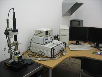
Nanoscope IIIa set |
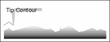
A surface image is constructed as a result of controlled movement of a special probe, which is called “tip” (thin sharpened wire; tip curvature radius is usually from several to tens of nm), keeping a constant distance from a surface. The line by line scanning tip movement reproduces a surface topography and a collected data is processed and displayed as an image on a computer screen. In order to keep a constant distance between a tip and a conducting sample surface, a phenomenon known as tunneling is used. If a potential difference is applied between a tip and a sample, while the tip is approached to a distance of several angstroms from the sample surface, a tunneling current starts to flow between them (this is a quantum effect and can only be observed on a micro-scale). The magnitude of tunneling current depends exponentially on a distance. For this reason, a tip movement across the surface is essentially performed in a constant current mode.
Basis of STM operation
Other similar technique is Atomic Force Microscopy (AFM). It also can achieve atomic resolution and its main advantage is that it can be used on all kinds of solid surfaces - both conducting and non-conducting. There are many types of AFM, whereas two most often used are Contact and Tapping Mode AFM. In Contact Mode AFM the image is obtained by scanning a sample surface with a cantilever. A cantilever approaches the surface up to the moment when it starts to bend. At this point it is moved across the surface in a way to keep its bending constant. Next, a cantilever movement data is processed (just like in STM) to obtain a digital image.

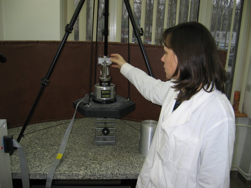
AFM microscope head
In Tapping Mode AFM, however, a cantilever is not directly moved across a surface, but is subjected to oscillation. It’s magnitude depends on a distance between a cantilever and a sample surface. Thus, a cantilever is moved across a surface in such a way, that its oscillation amplitude remains constant. The advantage of Tapping Mode AFM is that a cantilever is not in a constant contact with a studied surface (the surface is only “touched” with a frequency of cantilever oscillation), which saves it from possible damage. The technique is used for studying of “soft” samples such as cell membranes and organelle.
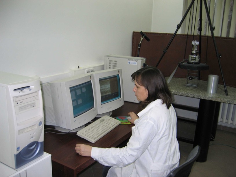
In our lab an integrated STM/AFM microscope is operated DI Nanoscope IIIa, Nanoscope V (Bruker) and XE Park Systems. It is usually used for imaging of metallic and organic nanostructures, polymer layers, polycarbonate membranes, tracking of liposome fusion etc.
Nanoscope V set-up